


Cell Biology Slides - 8/10/99 - 1 of 3
|
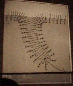
1) Topology of a plasma membrane when pulled into an aqueous region |
|
2)Villi of the small intestine - Epithelia (Outer layer) vs. Mesenchym(inner cells) |
|
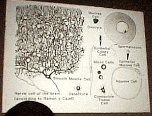
3) Different cells demonstrating all cell have a plasma membrane |
|
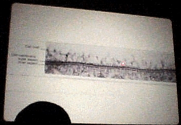
4) Stained cells viewed under light Microscope (Do you see PM? NO) |
|
5) EM of plasma membrane - Glycocalyx - sugar coat surrounding cell |
|
6) Polar head groups give the membrane a charge potential |
|
7) Cholesterol used to fill the leaks of the PM |
|
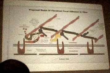
8) Peripheral protein network of cells |
|
9) Plasma membrane EM-sugar moieties not internal to the membrane |