 |
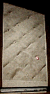
1) Low EM of Limb Bud Mesenchyme |
 |
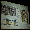
2) Lower level = Hypertrophied cells. They are mineralized
and secreting type 10 collagen Upper cells are chondroclasts (Hylarcartilage
take a columnar appearance) |
 |
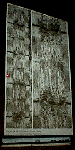
3) Bottom: Bone marrow of the Epiphysis, Bone matrix of
the epiphysis, zone of resting cartilage, proliferating cartilage, maturing
cartilage, calcifying cartilage, ? (In respective order from bottom to
top) |
 |
4) EM of the Muscle - Extracted with glycerol - see the
individual myofibrils - the fibrils are lined up in register |
 |
5) Thin section of a muscle = myofibrils are composed of
finer filaments |
 |
6) 200 angstroms - Clearly see the filaments that compose
the myofibril. See the thick and thin filament. Thick filaments have branches
that reach out to the thin filaments. |
 |
7) Myosin in solution = Kept from aggrigrating by using
high salt. See the tail and one or two heads |
 |
8) Self assembly of thick filaments that form by insoluble
tail regions - They are Bipolar |
 |
9) See the helical structure and the G subunits |
 |
10) Thin filaments decorated with myosin head groups vs.
non-attatched |
 |
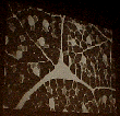
11) Neuromuscular Junction |
 |
12) Surface of the sarcolemma (They also showed a cross
section of the sarcolemma, but I missed the picture ) |
 |
13) Shows T tubule and sarcoplasmic recticulum |
 |
14) Depolarization signals the release the release of calcium
causes conformational change. It then triggers a release of Ca. |
 |
15) Fibroheterogeneity- Some fibers stain differently |
 |
16) Satillite Cells - = A myoblast - it sits beneath the
basement lamina and sarcolemma. |