Cell Biology 9/7/99
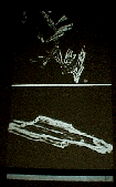 |
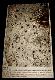
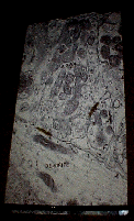
1) Isolated cardiac cells prepared with trypsin and hydrogenase |
 |
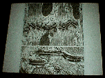
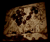
2) Thin section of cardiac muscle. See the intercalating disks. They myofibrils are branched. Big grey patches are the mitochondria. Nucleus in the upper left corner. |
 |
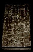
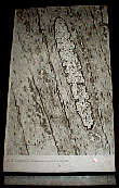
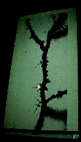
3) Intercalating Disks - Thinner slice of cardiac muscle. |
 |
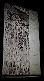
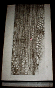
4) Gap Junctions |
 |
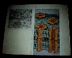
5) Myocytes - Lower middle = FA = Adherins junctions MA = Macula adherins N = Gap Junctions = Nexis junctions on lateral surfaces or between the FA or MA junction. |
 |
6) Thyroxin can switch on different sets fo contractile => faster contractile proteins => Cardiac skeletal type heterogeneity |
 |
7) Smooth Muscle |
 |
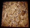
8) Thick, thin and microvessels |
 |
9) Again see the filaments - Thick => myosin that exist in filamentous state when it contracts Thin => Activated by increased cytosolic Calcium, They function in sliding filament action Intermediate => Interconnected dense bodies |
 |
10) SEM of Relaxed Smooth Muscle |
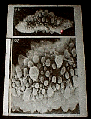 |
11) SEM of Contracted Smooth Muscle |
 |
12) Neuron- See Nissle body, RER, Nucleus (Very euchromatic), Lysosomes |
 |
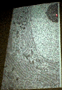
13) Neuropil |
 |
14) Synaptic spine or Dendritic Knobs |
 |
15) Neuromuscular junction synapse |
 |
16) Motor Neuron |
 |
17) Synapse in a central nervous system |